

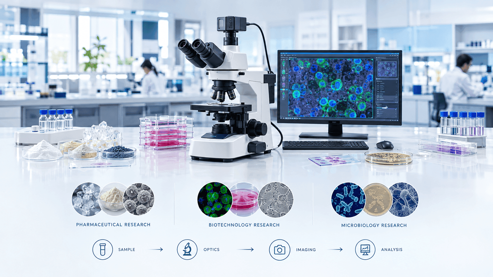
Microscopy plays a critical role in pharmaceutical, biotechnology and microbiology research. From examining microorganisms and cultured cells to evaluating formulations, crystals, particles and biological structures, the right microscope can turn an otherwise difficult observation into reliable visual information.
However, choosing a microscope for a research laboratory is not simply about selecting the highest magnification.
Different applications require different combinations of resolution, contrast, illumination, optical configuration, imaging capability, sample handling and analysis software.
A microbiology laboratory may prioritize phase contrast or fluorescence for observing microorganisms. A cell-biology research group may need an inverted microscope for cultured cells. A pharmaceutical laboratory may require polarized-light microscopy for crystals or specialized imaging for particulate contamination and formulation analysis.
The correct approach is therefore to start with the sample and research question, then select the microscope configuration that can answer it.
Why Microscope Selection Matters in Pharmaceutical and Biotech Research
Research microscopy is often used to answer specific scientific or quality-related questions.
Examples include:
Is a microorganism present?
What is its morphology?
How does a cell respond to a treatment?
Are particles present in a formulation?
What is the morphology of a pharmaceutical crystal?
How are cells distributed within a sample?
Are specific structures expressing a fluorescent marker?
Has a biological culture changed over time?
What does a material or formulation look like at microscopic scale?
A microscope that is appropriate for one of these applications may be poorly suited to another.
For example, a high-magnification brightfield system may be excellent for stained specimens but inadequate for live, unstained cells.
Similarly, a fluorescence-capable microscope may be unnecessary for routine brightfield teaching or basic sample inspection.
The goal is not maximum specification.
The goal is application-appropriate performance.
Microscope Selection by Research Application
A useful starting point is to map common research applications to microscope types.
Research Application | Commonly Suitable Microscope Approach |
|---|---|
Routine microbiology | Compound light microscope |
Stained microorganisms | Brightfield microscope |
Live microorganisms | Phase-contrast or other suitable contrast methods |
Cell culture | Inverted microscope |
Fluorescently labelled cells | Fluorescence microscope |
Pharmaceutical crystals | Polarizing microscope |
Particulate inspection | Brightfield/darkfield or specialized imaging |
Opaque materials | Reflected-light microscope |
Surface morphology | Stereo or materials microscope |
Advanced cellular imaging | Fluorescence/confocal or other specialized platform |
High-resolution ultrastructure | Electron microscopy |
These are starting points rather than universal rules.
The exact configuration should be determined by the sample, required resolution, imaging method and research workflow.
Step 1: Define the Research Question
Before comparing microscope brands or specifications, define what the laboratory needs to accomplish.
Ask:
What is the specimen?
Is it:
Bacteria?
Fungi?
Parasites?
Mammalian cells?
Tissue?
Pharmaceutical crystals?
Powder?
Particles?
Biological material?
Opaque material?
Is the sample alive?
Live samples often require different observation methods from fixed or stained specimens.
Is staining possible?
If the sample can be stained, conventional brightfield microscopy may provide sufficient contrast.
If staining is undesirable or impossible, phase contrast, fluorescence or other contrast techniques may be more appropriate.
What level of detail is required?
Determine whether the research requires:
General morphology
Cellular structures
Subcellular features
Particle characteristics
Quantitative measurements
Time-lapse observations
Are images required?
If images will be used for publications, reports, quality investigations or quantitative analysis, digital imaging should be considered from the beginning.
Step 2: Choose Between Upright and Inverted Microscopes
One of the first configuration decisions is whether the laboratory needs an upright or inverted microscope.
Upright Microscopes
In an upright microscope, the objectives are positioned above the specimen.
They are commonly used for:
Prepared slides
Tissue sections
Stained microbiological samples
Histology
Routine microbiology
Fixed specimens
For laboratories that primarily work with slides, an upright microscope is often a practical starting point.
Inverted Microscopes
In an inverted microscope, the objectives are positioned below the specimen stage.
This configuration is particularly useful for specimens in containers such as culture dishes or plates.
Common applications include:
Cell culture
Live-cell research
Tissue culture
Biotechnology research
Drug-response studies
An inverted microscope can allow researchers to observe cells without repeatedly transferring them to conventional microscope slides.
For pharmaceutical and biotechnology laboratories performing cell-based assays, this can be an important consideration.
Step 3: Understand the Role of Brightfield Microscopy
Brightfield is one of the most widely used microscopy techniques.
It is particularly effective when specimens provide sufficient contrast, either naturally or through staining.
Common applications include:
Stained bacteria
Fungal structures
Tissue sections
Histological samples
Pharmaceutical preparations
General morphology
For routine microbiology, a high-quality brightfield microscope with appropriate objectives can be extremely useful.
The limitation is that transparent, unstained specimens may provide little contrast.
That is where alternative techniques become important.
Step 4: When Should You Choose Phase Contrast?
Phase contrast is designed to improve the visibility of transparent specimens that have relatively low contrast under conventional brightfield illumination.
It can be particularly useful for:
Live cells
Cell cultures
Unstained microorganisms
Biological specimens
For biotechnology research, phase contrast can make it easier to monitor changes in:
Cell morphology
Cell density
Attachment
Growth
General culture condition
If the research workflow involves frequent observation of live cultured cells, phase contrast may be more useful than simply purchasing higher-magnification objectives.
Step 5: When Is Fluorescence Microscopy Needed?
Fluorescence microscopy is valuable when researchers need to detect specific fluorescent signals.
Fluorescent labels can allow researchers to visualize particular structures, molecules or organisms against a contrasting background.
Applications can include:
Immunofluorescence
Microbial identification research
Cellular localization studies
Molecular biology
Drug-response research
Biomarker studies
A fluorescence microscope typically requires more than simply adding a fluorescence light source.
The optical system may need:
Appropriate excitation illumination
Excitation filters
Dichroic mirrors
Emission filters
Suitable objectives
Sensitive camera
Imaging software
The required filter sets should be based on the fluorophores used by the laboratory.
Step 6: Consider Confocal Microscopy for Advanced Research
For some biotechnology and pharmaceutical research applications, conventional fluorescence microscopy may not provide sufficient optical sectioning.
Confocal microscopy can improve optical sectioning and support imaging of structures within thicker specimens.
Potential applications include:
Three-dimensional cellular imaging
Tissue imaging
Fluorescent localization
Multichannel imaging
Advanced cell biology
However, confocal microscopy is considerably more specialized and expensive than a conventional research microscope.
A laboratory should therefore establish a clear scientific requirement before investing in such a system.
If the required work can be performed adequately using widefield fluorescence, a simpler system may provide better value.
Step 7: Pharmaceutical Research and Polarized-Light Microscopy
Polarized-light microscopy can be particularly useful in pharmaceutical research where the optical properties of crystals or other anisotropic materials are important.
Potential applications include:
Crystal morphology
Crystalline materials
Solid-state characterization support
Particle examination
Formulation research
Certain pharmaceutical materials can exhibit characteristic behavior under polarized light.
A polarizing microscope can therefore provide information that ordinary brightfield observation may not reveal.
However, microscopy should generally be considered as one analytical tool within a broader pharmaceutical characterization workflow.
Step 8: Microscopy for Pharmaceutical Particle and Contamination Analysis
Pharmaceutical laboratories may need to inspect particles or foreign material.
The research or quality-control question may involve:
Particle presence
Particle morphology
Particle size estimation
Shape
Distribution
Foreign material identification support
Depending on the application, laboratories may consider:
Brightfield
Darkfield
Polarized illumination
Digital imaging
Automated image analysis
For quantitative particle analysis, microscope optics and image-analysis software should be evaluated together.
Simply capturing a high-resolution image does not automatically provide reliable measurements.
Calibration and appropriate analysis methods are equally important.
Step 9: Microbiology Research Requirements
Microbiology laboratories work with organisms that can vary significantly in size, morphology and optical properties.
The microscope should therefore match the organisms and methods used.
Bacteria
Routine bacterial morphology may be observed using:
Brightfield
Oil-immersion objectives
Phase contrast
Fluorescence
Fungi
Fungal structures may require:
Brightfield
Phase contrast
Specialized staining
Fluorescence in research applications
Parasites
Depending on the organism, microscopy may involve:
Brightfield
Phase contrast
Specialized staining
Fluorescence
Live Microorganisms
For living organisms, phase contrast or other suitable contrast methods can provide improved visibility without relying exclusively on staining.
Step 10: Numerical Aperture Matters More Than Magnification Alone
One of the most important microscope-selection principles is:
Higher magnification does not automatically mean higher resolution.
Resolution is influenced by the optical system, including numerical aperture, wavelength and other factors.
For example, a high-quality 40× objective can provide more useful information than a poorly designed objective offering a much higher nominal magnification.
When evaluating objectives, consider:
Magnification
Numerical aperture
Working distance
Optical correction
Immersion requirements
Compatibility with the microscope system
Research laboratories should evaluate objective specifications as part of the complete optical system.
Step 11: Understand Working Distance
Working distance is the distance between the objective's front lens and the specimen when the specimen is in focus.
A shorter working distance can accompany high-resolution objectives, while specialized applications may benefit from longer working distances.
Longer working distance can be valuable when:
Samples are relatively thick
Containers are involved
Manipulation is required
Specialized experimental setups are used
Therefore, the objective with the highest numerical aperture is not automatically the best choice for every experiment.
Step 12: Select the Right Contrast Method
A pharmaceutical, biotechnology or microbiology laboratory may need more than brightfield.
Common techniques include:
Brightfield
Best suited to specimens with adequate natural or stained contrast.
Phase Contrast
Useful for transparent, low-contrast live specimens.
Darkfield
Can improve visibility of certain small or low-contrast specimens.
Fluorescence
Useful for detecting fluorescently labelled structures.
Polarized Light
Useful for examining materials with relevant optical anisotropy.
Differential Interference Contrast
Can provide enhanced contrast and relief-like appearance for suitable specimens.
The laboratory should choose techniques based on actual research requirements rather than attempting to maximize the number of features.
Step 13: Evaluate Digital Imaging Carefully
For modern research laboratories, the camera is often as important as the microscope.
A digital imaging system should be evaluated for:
Sensor technology
Resolution
Pixel size
Sensitivity
Dynamic range
Frame rate
Exposure control
Field of view
Interface
Software integration
Different experiments require different camera characteristics.
For live-cell imaging, frame rate can be important.
For fluorescence, sensitivity and signal-to-noise performance may be more important.
For documentation and routine morphology, resolution and color reproduction may receive greater emphasis.
Step 14: Consider Image Analysis
If the laboratory needs quantitative information, image analysis becomes a major part of the microscope workflow.
Possible applications include:
Cell counting
Particle counting
Size measurement
Area measurement
Morphological analysis
Fluorescence intensity analysis
Cell viability assessment
Colony or structure analysis
The system should support appropriate calibration and reproducible measurement procedures.
A research laboratory should also consider whether image-analysis workflows can be exported and documented for future review.
Step 15: Think About Reproducibility
Research microscopy should produce observations that can be reproduced.
This becomes especially important when images form part of scientific publications or quantitative studies.
Consider:
Consistent illumination
Stable focus
Appropriate calibration
Consistent exposure
Objective identification
Standardized acquisition settings
Proper image storage
Metadata retention
Automated acquisition can sometimes help standardize imaging conditions, but automation should be adopted when it solves a real workflow problem.
Step 16: Consider Live-Cell Research Requirements
Biotechnology and pharmaceutical research frequently involves living cells.
A live-cell microscopy system may require more than an optical microscope.
Depending on the experiment, researchers may need:
Inverted configuration
Environmental control
Temperature stability
CO₂ control
Humidity management
Appropriate culture vessels
Low-phototoxicity imaging
Time-lapse acquisition
Automated stage movement
Not every cell-culture laboratory needs all of these features.
The correct configuration depends on whether the work involves brief observations or long-duration live-cell experiments.
Step 17: Evaluate Phototoxicity and Photobleaching
Fluorescence-based live-cell research introduces another consideration.
Repeated or intense illumination can affect fluorescent signals and, depending on the specimen and conditions, can influence biological samples.
Researchers should therefore consider:
Illumination intensity
Exposure time
Acquisition frequency
Fluorophore characteristics
Imaging duration
Sample sensitivity
For experiments requiring long time-lapse imaging, minimizing unnecessary illumination can be important.
Step 18: Ergonomics and Laboratory Workflow
Research scientists can spend many hours at a microscope.
Ergonomics therefore affects both comfort and productivity.
Important considerations include:
Eyepiece height
Viewing angle
Smooth focusing
Stage control position
Objective switching
Camera placement
Seating position
Control accessibility
If the microscope is used primarily through a monitor rather than eyepieces, the workstation should also be designed around the digital workflow.
Step 19: Software and Data Management
Modern research microscopes can generate large amounts of image data.
Before purchasing a system, consider:
Image file formats
Storage requirements
Software licensing
Analysis capabilities
Export options
Data backup
Compatibility with laboratory systems
Long-term accessibility
A microscope that creates excellent images but locks researchers into an inconvenient data workflow can create problems later.
For research environments, open and well-documented data workflows can be valuable.
Step 20: Maintenance and Service Support
A research microscope is a precision instrument.
Routine maintenance and professional service can affect its performance over time.
Before purchase, evaluate:
Warranty
Preventive maintenance
Local service availability
Replacement parts
Objective servicing
Camera support
Software updates
Technical training
Expected service response time
For pharmaceutical and biotechnology laboratories, downtime can affect experiments and project schedules.
Service support should therefore be treated as part of the purchase decision rather than an afterthought.
Research Microscope vs Routine Laboratory Microscope
Not every laboratory needs a premium research platform.
Requirement | Routine Laboratory | Research Laboratory |
|---|---|---|
Basic morphology | High priority | High priority |
Advanced optics | Sometimes unnecessary | Often important |
Phase contrast | Application-dependent | Frequently useful |
Fluorescence | Application-dependent | Common in relevant research |
Digital imaging | Useful | Often essential |
Image analysis | Limited/basic | Frequently important |
Automation | Usually limited | Application-dependent |
Live-cell capability | Basic/optional | Important for relevant research |
Upgradeability | Useful | Highly valuable |
Data management | Basic | Important |
Service | Important | Critical |
The best procurement strategy may involve multiple microscope configurations rather than one universal platform.
How to Select a Microscope for a Pharmaceutical Laboratory
A pharmaceutical research laboratory should first identify its primary applications.
If the focus is formulation and particle observation:
Consider:
Brightfield
Polarized light
Digital imaging
Appropriate measurement software
If the focus is cell-based drug research:
Consider:
Inverted microscope
Phase contrast
Fluorescence
Digital imaging
Time-lapse capability where required
If the focus is microbiological research:
Consider:
High-quality compound optics
Appropriate objectives
Phase contrast
Fluorescence where needed
Digital documentation
If the focus is advanced cellular research:
Consider:
Fluorescence
Confocal capability
Automated acquisition
Image analysis
Environmental control where required
How to Select a Microscope for Biotechnology Research
Biotechnology applications can range from routine cell culture to highly specialized molecular and cellular imaging.
A useful selection framework is:
Sample → Contrast → Resolution → Imaging → Analysis
For example:
Live cultured cells
↓
Phase contrast
↓
Appropriate objective
↓
Sensitive digital camera
↓
Cell morphology and quantitative analysis
This approach prevents laboratories from buying expensive features that do not contribute to the intended experiment.
How to Select a Microscope for Microbiology Research
For microbiology, consider:
Organism Size
Smaller organisms may require higher-resolution objectives and appropriate contrast.
Sample Preparation
Determine whether samples are stained, fixed or live.
Imaging Requirements
Will the laboratory simply observe morphology or capture images for analysis?
Fluorescence
If fluorescent markers are used, ensure the microscope supports the required excitation and emission ranges.
Oil Immersion
For certain high-resolution applications, an oil-immersion objective may be appropriate.
Digital Documentation
If images need to be archived or analyzed, include camera and software requirements in the procurement specification.
Common Mistakes When Choosing a Research Microscope
Buying Based on Magnification Alone
High magnification does not guarantee useful resolution.
Choosing a Microscope Before Defining the Sample
The specimen should determine the optical configuration.
Ignoring Objectives
Objectives are central to microscope performance.
Treating Cameras as Accessories
For imaging-heavy research, camera performance can directly affect experimental results.
Buying Every Available Contrast Technique
Advanced features are valuable only when they solve a real research problem.
Ignoring Software
Image acquisition and analysis are increasingly integrated with microscopy workflows.
Forgetting Future Applications
Research programs evolve. Upgradeability can extend the useful life of the system.
Ignoring Service Availability
A microscope that cannot be maintained efficiently can become a costly laboratory bottleneck.
A Practical Microscope Selection Checklist
Before purchasing a microscope for pharmaceutical, biotechnology or microbiology research, answer the following.
Sample
What specimens will be examined?
Are they live, fixed or stained?
Are they transparent or opaque?
What is their approximate size?
Optical Requirements
What resolution is required?
What magnification range is needed?
What numerical aperture is appropriate?
What working distance is required?
Contrast
Brightfield?
Phase contrast?
Darkfield?
Fluorescence?
Polarization?
DIC?
Imaging
Is a camera required?
What sensitivity is needed?
Is time-lapse imaging required?
Is quantitative analysis required?
Workflow
Upright or inverted?
Manual or automated?
Will multiple researchers share the system?
What sample holders or culture vessels are used?
Data
What software is needed?
What measurements must be performed?
How will images be stored?
What file formats are required?
Lifecycle
What is the warranty?
Where is technical support available?
What are the maintenance requirements?
Can the system be upgraded?
Total Cost of Ownership
The microscope's purchase price is only one part of the investment.
A realistic procurement assessment should consider:
Microscope
Objectives
Illumination
Camera
Filters
Specialized accessories
Software
Installation
Training
Maintenance
Upgrades
=
Total Cost of Ownership
A low-cost microscope may not remain economical if it requires frequent repairs or cannot support future research applications.
At the same time, an advanced research platform can be an inefficient investment if the laboratory only performs routine brightfield observations.
The objective should be fit-for-purpose performance over the instrument's expected service life.
Should You Buy One Microscope for Every Application?
Usually, not necessarily.
A pharmaceutical or biotechnology organization may benefit from a combination of systems.
For example:
Routine microbiology
→ Compound brightfield microscope
Cell culture
→ Inverted phase-contrast microscope
Fluorescence research
→ Fluorescence-capable imaging system
Crystal analysis
→ Polarizing microscope
Advanced cellular imaging
→ Confocal or other specialized platform
This approach allows each instrument to be optimized for its intended role.
It can also prevent an expensive research microscope from becoming overloaded with routine work.
Final Verdict: How to Choose the Right Research Microscope
The right microscope for pharmaceutical, biotechnology and microbiology research is determined by the scientific question, sample characteristics and imaging workflow.
For routine microbiology, high-quality compound optics and appropriate objectives may be sufficient.
For live-cell biotechnology research, an inverted microscope with suitable contrast methods may be more appropriate.
For fluorescently labelled specimens, fluorescence capability and appropriate filters and imaging hardware become essential.
For pharmaceutical crystal or formulation research, polarized-light microscopy can provide valuable additional information.
For advanced cellular imaging, fluorescence, confocal or automated systems may be justified when the research question requires them.
The most effective selection process follows a simple chain:
Application → Sample → Contrast → Resolution → Imaging → Analysis → Workflow → Lifecycle Cost
Instead of asking:
"Which microscope has the highest specifications?"
ask:
"Which microscope can produce the information our research actually requires, reliably and reproducibly?"
That question leads to a much more defensible laboratory-equipment decision.