

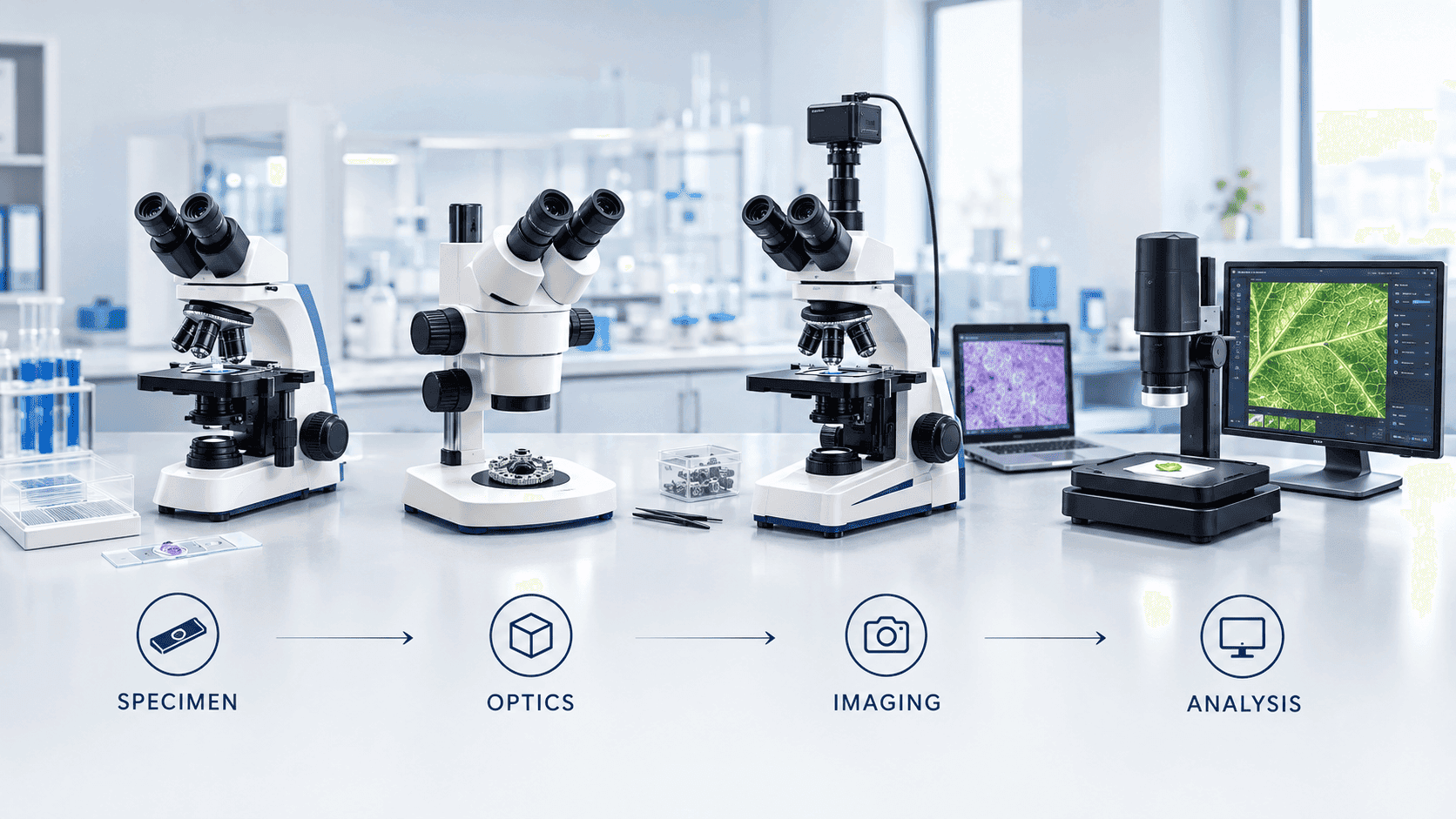
Choosing a microscope becomes much easier when you stop starting with magnification. The better starting point is the specimen, the information you need from it, and the way the image will be used.
A compound microscope, stereo microscope, trinocular microscope and digital microscope are not simply four competing versions of the same instrument. They describe different aspects of microscopy: optical design, viewing geometry, camera integration and image delivery. One microscope can, for example, be compound, trinocular and digital at the same time.
Current microscopy guidance similarly distinguishes compound, stereo and digital systems by optical design and highlights the trade-off between field of view and resolution.
The short answer: which microscope should you choose?
Main task | Starting point | Why |
Cells, tissues, bacteria or prepared slides | Compound microscope | Designed for fine detail in thin specimens. |
Dissection, insects, plants, minerals or components | Stereo microscope | Wide field, working distance and stereoscopic depth. |
Direct viewing plus permanent camera integration | Trinocular microscope | Dedicated camera path while retaining eyepiece observation. |
Screen-based viewing, documentation or collaboration | Digital microscope / imaging system | Camera, display and software-centered workflow. |
Advanced optical viewing plus imaging | Trinocular compound or stereo + camera | Combines direct observation with dedicated imaging. |
1. Compound microscopes: when fine detail matters
Compound microscopes are designed for higher-magnification observation of relatively small specimens, commonly prepared on slides. They are widely used for cell biology, microbiology, histology, pathology education and laboratory research.
They generally use higher-NA objective lenses and provide a comparatively smaller field of view and shorter working distance than stereo systems. Educational microscopy guidance contrasts compound systems with stereoscopic instruments on specimen size, magnification, field of view and working distance.
Typical applications:
Cells and tissues
Microorganisms and prepared microbiology slides
Histology and pathology specimens
Thin plant sections
Fluorescence or phase-contrast work when suitably configured
Research requiring fine optical detail
2. Stereo microscopes: when the whole specimen matters
A stereo microscope, also called a dissecting microscope, uses separate optical paths to provide stereoscopic depth perception. It is particularly useful for larger, solid specimens, surface inspection and tasks where the user needs room to manipulate the object.
University bioimaging guidance describes stereo microscopy as a reflection-based approach suited to surface visualization, low magnification and specimen manipulation.
Dissection and anatomy teaching
Insects and biological specimens
Leaves, flowers and whole organisms
Minerals and geological samples
Electronics and component inspection
Surface defects and manufacturing inspection
Tasks requiring tools beneath the objective
3. Trinocular microscopes: when viewing and imaging must work together
Trinocular does not describe a completely different application class. It describes an observation head with a dedicated third optical path or camera port. A trinocular compound microscope remains a compound microscope; a trinocular stereo microscope remains a stereo microscope.
The practical benefit is workflow flexibility: the user can observe through the eyepieces while a camera is connected for documentation, teaching, image capture or analysis. A trinocular configuration is particularly useful when imaging is routine rather than occasional.
Permanent camera installation
Direct optical observation plus camera output
Classroom display and demonstrations
Repeatable research image acquisition
Routine documentation and measurement
4. Digital microscopes: when the image needs to live on a screen
Digital microscopy shifts the workflow toward a camera and display. Depending on the design, the optical system may use fixed or zoom optics and may be based on compound or stereo architecture. Leica notes that digital microscopes can use either fixed or zoom optics; fixed optics generally favor resolution while zoom systems favor a larger field of view.
Digital systems can be especially useful when images must be shared, captured, measured, annotated, recorded or reviewed by multiple people. They are common in education, inspection, documentation and collaborative laboratory workflows.
Compound vs. stereo microscope: the key differences
Factor | Compound | Stereo |
Specimen | Thin, small, often slide-mounted | Larger, solid, intact or opaque |
Lighting | Often transmitted | Often reflected/incident; transmitted options also exist |
Magnification | Higher | Low to medium |
Field of view | Smaller | Larger |
Working distance | Shorter | Longer |
Depth perception | Not true stereoscopic viewing | True stereoscopic viewing |
Manipulation | Limited | Much easier |
Typical use | Cells, tissues, microbes, fine structures | Dissection, morphology, surfaces, components |
Trinocular vs. digital: not actually opposites
A trinocular microscope provides a dedicated camera path while retaining optical observation. A digital microscope emphasizes camera-and-display imaging. Therefore, a trinocular microscope can be part of a digital imaging setup.
Question | Trinocular | Digital |
Eyepiece viewing | Yes | Depends on design |
Camera connection | Core capability | Usually integrated/direct |
Best fit | Optical + imaging workflow | Display-first workflow |
Teaching display | Strong with monitor/projector | Strong |
Research imaging | Strong with appropriate camera | Strong when sensor/software fit |
Upgrade flexibility | Often high | Depends on architecture |
Which microscope fits each application?
Application | Starting point | Key consideration |
Biology teaching | Compound | Slide specimens and ease of use. |
Microbiology | Compound | Resolution, objectives and contrast options. |
Dissection / zoology | Stereo | Working distance and depth perception. |
Botany / morphology | Stereo + compound | Whole specimens vs cellular sections. |
Histology | Compound | Objectives and optical quality. |
Cell culture | Compound with suitable contrast | Transparent live cells need appropriate contrast. |
Fluorescence research | Compound fluorescence platform | Filters/channels, sensitivity and camera. |
Electronics inspection | Stereo or digital stereo | Working distance, field and surface lighting. |
Materials inspection | Application-specific system | Surface geometry and resolution. |
Classroom demonstration | Digital or trinocular + camera | Display and capture workflow. |
Measurement/documentation | Digital or trinocular imaging | Camera, calibration and software. |
Specifications that actually matter
Magnification
Do not buy based on the largest number. Useful magnification depends on the optical system and resolving power.
Resolution and numerical aperture
Objective quality and numerical aperture are critical for fine-detail work.
Working distance
Especially important for stereo, dissection and inspection because it determines usable space beneath the objective.
Field of view
A wider field helps with larger specimens; a smaller field can suit fine cellular detail.
Illumination
Match transmitted, reflected or specialized illumination to the specimen.
Camera
Evaluate sensitivity, pixel size, frame rate, dynamic range and field of view not megapixels alone.
Software
If measurement, annotation, stitching, time-lapse or documentation is required, software becomes part of the microscope workflow.
Ergonomics
Consider viewing angle, focusing, stage movement and user comfort.
Service and upgradeability
Evaluate warranty, local support, replacement parts, training and future expansion.
Common microscope-buying mistakes
Choosing the highest advertised magnification instead of matching the specimen.
Confusing binocular with stereo: two eyepieces do not automatically create stereoscopic viewing.
Treating trinocular as a separate microscope type rather than an imaging configuration.
Assuming every digital microscope has the optical performance of a research compound microscope.
Buying a camera without considering objectives, field of view and software.
Using a stereo microscope where cellular-level resolution is required.
Using a compound microscope for large opaque specimens requiring manipulation.
Ignoring working distance and illumination.
Comparing cameras by megapixels alone.
Overlooking service coverage and lifecycle cost.
A practical microscope decision tree
1. Thin specimen on a slide? → Start with a compound microscope.
2. Larger, solid or opaque specimen? → Start with a stereo microscope.
3. Need true depth perception or space for manipulation? → Stereo.
4. Need cellular or microbial detail? → Compound.
5. Need a permanent camera path plus eyepiece observation? → Trinocular.
6. Need screen-based viewing, documentation or software analysis? → Digital imaging.
7. Need both direct viewing and digital imaging? → Consider a trinocular system with a suitable camera.
8. Need fluorescence, phase contrast or another specialized method? → Specify the optical platform and contrast method together.
The right answer may be a combination
Many laboratories do not need to choose a single category. A university biology department may use compound microscopes for cells and microorganisms and stereo microscopes for dissection and morphology. A research laboratory may add a trinocular head and camera to either platform. A teaching laboratory may prioritize digital output so one specimen can be displayed to an entire class.
The most reliable procurement sequence is: define the application → identify the specimen → choose the optical architecture → determine the imaging workflow → compare specifications → validate service and lifecycle cost.
Final takeaway
Compound, stereo, trinocular and digital microscopes describe different parts of the microscopy decision. Compound and stereo primarily describe optical approaches. Trinocular describes a camera-ready observation configuration. Digital describes an image-centered workflow.
If the specimen is thin and microscopic, start with compound. If it is larger, solid and three-dimensional, start with stereo. If imaging must happen alongside direct observation, consider trinocular. If collaboration, documentation or software analysis is central, consider digital imaging. The right microscope is the one that produces the information your application actually requires.