

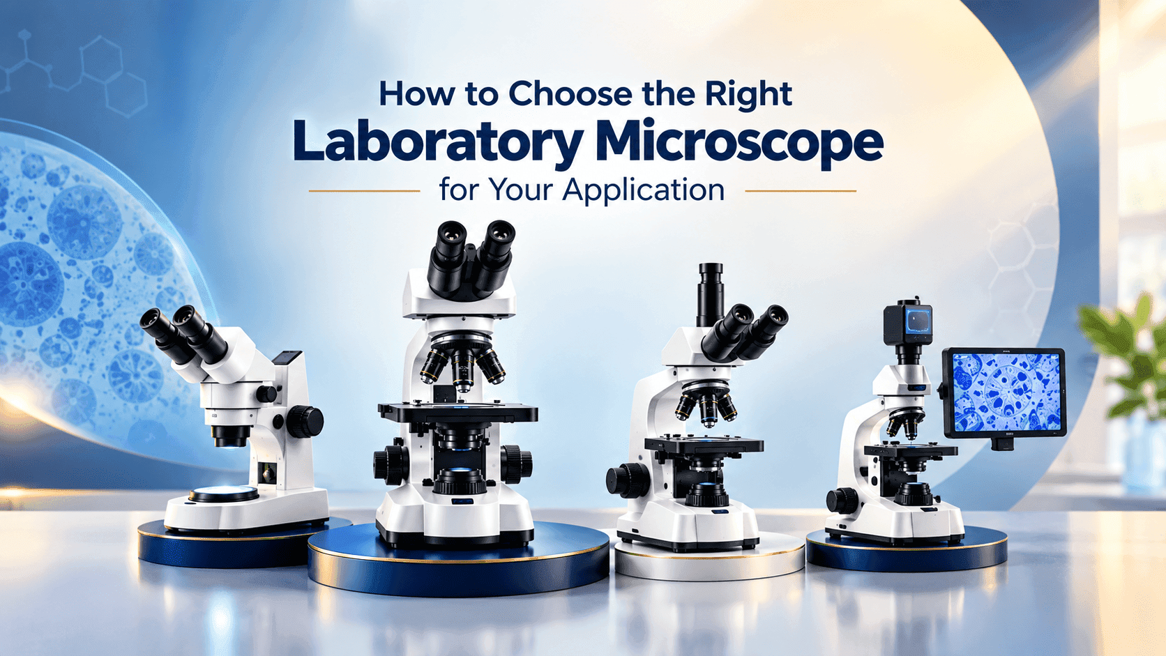
Start With the Application, Not the Microscope
The right laboratory microscope is not necessarily the one with the highest magnification, the most accessories, or the highest price. It is the system that produces the information your application needs with appropriate optics, contrast, illumination, sample access, imaging, and ergonomics.
Start by answering: What do you need to see, measure, document, compare, or manipulate? similarly emphasizes specimen type, application, contrast, documentation, spatial dimensions, and user requirements.
What specimens will be examined cells, tissue, microorganisms, plants, materials, components, powders, or larger objects?
Are specimens fixed, stained, unstained, living, fluorescent, reflective, opaque, or transparent?
Is the workflow observation only, or does it require photography, measurement, image analysis, reporting, teaching, or publication?
Will samples be on slides, in culture dishes, flasks, chambers, or multiwell plates?
Is the workflow routine and repetitive, or does it need fluorescence, automation, multidimensional imaging, or future expansion?
3. Match Microscope Type to the Job
Type | Typical use | Strength | Watch-out |
Compound upright | Slides, stained tissue, microbiology, routine biology | High-resolution transmitted-light work | Less convenient for large culture vessels |
Inverted | Cell culture, live cells, dishes, flasks, plates | Easy access to culture vessels | Must match objectives and stage to vessel geometry |
Stereo / dissecting | Dissection, sorting, inspection, larger 3D objects | Large field of view, depth perception, long working distance | Lower resolution than compound systems |
Digital | Documentation, inspection, collaboration, measurement | Monitor-based viewing and image capture | Camera, optics, software and display all matter |
Fluorescence | Fluorescent labels, proteins, biomarkers | Specific molecular/structural signals | Needs suitable filters, illumination, objectives and camera |
Research platform | Advanced life-science research | Expandable contrast, imaging and automation | Higher cost and configuration complexity |
compound systems as higher-resolution options with smaller fields of view, while stereo systems offer larger fields of view with lower resolution; digital systems can use fixed or zoom optics.
Upright vs. Inverted Microscopes
Specimen geometry often determines the choice. Upright microscopes are generally suited to slide-based work, while inverted microscopes place objectives below the stage and are particularly useful for specimens maintained in culture vessels.
inverted microscopes for routine examination of living cells, including morphology and confluency checks, with research configurations supporting fluorescence and live-cell work.
Histology/pathology slides → upright compound system.
Routine microbiology on prepared slides → upright compound with appropriate objectives/contrast.
Adherent cells in dishes or flasks → inverted microscope, often with phase contrast.
Live-cell time-lapse → inverted research platform with appropriate camera and environmental control.
Dissection or larger 3D samples → stereo microscope.
Measurement/QC → compound, stereo, or digital measurement system depending on feature size and geometry.
Resolution Matters More Than Magnification
Magnification enlarges an image; resolution determines whether fine structures can actually be distinguished. Excessive magnification without adequate resolution can create a larger image without adding useful information.
Numerical aperture (NA) is a major determinant of objective performance. Higher NA generally supports greater resolving power and light collection, although it comes with optical and working-distance trade-offs.
Choose Objectives as Carefully as the Microscope
Magnification - how large the specimen appears.
Numerical aperture - strongly related to resolution and light collection.
Working distance - the physical clearance between objective and specimen.
Immersion medium - air, oil, water or another medium, depending on objective design.
Optical correction - important for chromatic/spherical aberration and field flatness.
Coverslip requirements - critical for many high-NA objectives.
Long-working-distance options - important for thick vessels, chambers, manipulation and industrial samples.
working distance generally decreases as magnification and NA increase, while specialized long-working-distance objectives provide greater specimen clearance through optical trade-offs.
Select the Right Contrast Method
Method | Useful when | Typical applications |
Brightfield | Stained or naturally contrasting specimens | Histology, routine microbiology, teaching |
Phase contrast | Transparent, unstained specimens | Live cells, cell culture, microorganisms |
DIC | Unstained transparent structures | Live cells and biological specimens |
Fluorescence | Specific labeled structures | Immunofluorescence, molecular/cell biology |
Darkfield | Scattering structures | Selected microorganisms and specialized specimens |
Polarized light | Birefringent/anisotropic features | Crystals, minerals, fibers, selected materials |
phase contrast and DIC can improve visualization of unstained biological specimens that are difficult to see in brightfield; phase contrast uses specialized objectives and DIC requires appropriate prisms.
Illumination Can Change What You See
A microscope should be evaluated as an optical system. Illumination must match the specimen and contrast method.
Transmitted illumination supports many brightfield, phase-contrast and DIC workflows.
Fluorescence requires controlled excitation and emission pathways.
Reflected-light applications may need dedicated illumination geometry.
Uniformity, stability, intensity control, heat and maintenance affect practical performance.
LED sources can be attractive for many routine workflows because of long service life and low maintenance, but the appropriate source remains application-dependent.
Digital Imaging: Buy the Workflow, Not Just the Camera
A digital microscope system is more than a camera. Optical resolution, sensor characteristics, pixel size, field of view, exposure control, software, monitor, storage and analysis all contribute to the final result.
Routine documentation → prioritize simple, repeatable capture and file management.
Publication imaging → evaluate color fidelity, resolution, dynamic range, stitching and metadata.
Quantitative analysis → confirm calibration, measurement, segmentation and export capabilities.
Live-cell imaging → prioritize sensitivity, speed, exposure control and environmental compatibility.
Teaching/collaboration → consider field of view, monitor ergonomics and easy sharing.
specifically identify camera pixel size, optical quality, resolution, software and application needs as selection factors.
Field of View, Working Distance and Sample Access
Maximum magnification is not always the priority. Locating a region of interest, orienting on a large specimen, manipulating a sample, or inspecting a 3D object may require a larger field of view and longer working distance.
Identify zoom range, field of view, working distance, depth of field and ergonomics as important stereo-microscope selection factors.
Ergonomics Is a Technical Requirement
Adjustable viewing angle and eyepiece position.
Comfortable focus and stage controls.
Stable stand and suitable vibration control.
Easy access to frequently used functions.
Appropriate seating/standing position.
Simple switching between users and imaging modes.
Ergonomics matters particularly in laboratories where users spend long periods at the microscope.
Application-to-Microscope Guide
Application | Starting configuration | Priority criteria |
Histopathology | Upright compound | Plan objectives, brightfield, fluorescence if needed, camera and ergonomics |
Microbiology | Upright compound | Appropriate objectives, brightfield/phase contrast, immersion where protocol requires |
Cell culture | Inverted | Phase contrast, long working distance, compact footprint, fluorescence option |
Fluorescence research | Upright or inverted research | Filter sets, sensitive camera, objectives, channel count, software |
Biotechnology | Often inverted/research compound | Live-cell compatibility, fluorescence, automation and analysis |
University teaching | Compound or digital compound | Durability, ease of use, shared viewing and broad objective set |
Dissection | Stereo | Working distance, depth perception, zoom and illumination |
Materials/components | Stereo or reflected-light | Working distance, reflected illumination, polarization and measurement |
Measurement/QC | Compound, stereo or digital | Resolution, calibration, camera, optics and software |
Quick Decision Questions
Slides and routine transmitted-light work → upright compound.
Living cells in dishes/flasks/plates → inverted.
Large 3D objects, dissection or manipulation → stereo.
Specific labeled structures → fluorescence.
Unstained live-cell contrast → phase contrast or DIC.
Measurement/documentation → camera + calibration + software + suitable optics.
Automation, high-content or multidimensional imaging → research platform.